Each year, about one in 10,000 persons in the United States suffers a retinal detachment. If untreated, serious and sometimes total loss of vision in the affected eye can result. Today advanced techniques for surgery make it possible to restore vision in most cases, but the key is early detection and diagnosis.
The retina is a thin, transparent membrane which lines the back of the eye. Detachment of the retina occurs when it separates from the wall of the eye. This usually happens when the vitreous, a gelatin-like substance which fills the eye, collapses and sags forward as it softens with age.
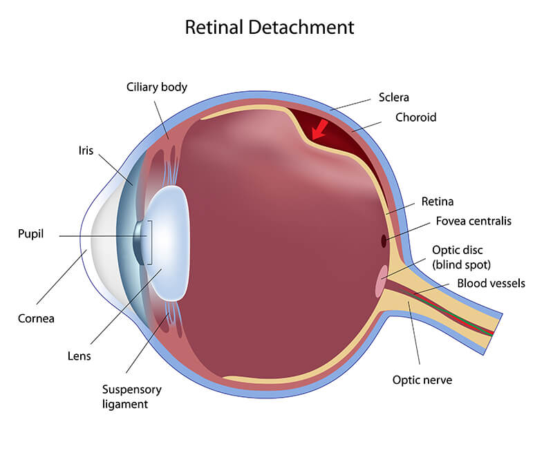
In the process it may pull on the retina and produce a tear or hole. Fluid from the vitreous can then leak through the retinal tear and detach the retina.
Retinal detachment is painless. Visual symptoms almost always appear before the retina detaches. As the vitreous shrinks and sags, it may tug on the retina and cause the sensation of flashing lights. If the tugging is strong enough and causes a retinal tear, small retinal blood vessels may be torn and the bleeding may cause the sudden appearance of new “floaters.” The sudden onset of flashing lights, especially in association with the sudden appearance of new “floaters” (dark or light spots or specks or lines in the field of vision) are possible warning symptoms of a retinal detachment. If this occurs, a physician eye specialist should be consulted immediately.
When a retinal tear or hole develops but has not yet progressed to retinal detachment, doctors can prevent detachment by sealing the area around the hole with laser or with a freezing device called the cryoprobe. The process is known as cryopexy. Neither laser nor cryopexy requires admission to a hospital, and can be performed on an outpatient basis.
If retinal detachment has occurred, it is perceived as a dark shadow or curtain moving over the eye; the larger the area of detachment, the larger the perceived shadow. If the central retina (the macula) becomes detached, reading and central vision are lost.
For retinal detachment, there are now several possible treatments for certain small detachments a recently developed gas bubble can be placed inside the eye which expands and flattens the retina against the wall of the eye. Laser or cryopexy is then applied to the tear. For more complicated detachments, admission to the hospital for surgery is frequently required, and an operation called a scleral buckle is typically performed. A scleral buckle is a piece of soft silicone which indents the outer wall of the eye, seals the tear and repairs the detachment. For yet other very complicated detachments, a microscopic intraocular procedure called vitrectomy is now performed.
Thanks to these advanced techniques, most retinal detachments can be repaired. If the detachment is not too extensive, there is a 90% chance that vision can be completely restored. Even in advanced detachments some useful vision can frequently be recovered and the retina physically reattached. The ultimate return of vision depends primarily on the severity and duration of the preceding retinal detachment. Early diagnosis and treatment are the best assurances for optimum return of vision.
For more information and a video tutorial please click on the instructional video below.
Colvard-Kandavel Eye Center offers personalized eye care services for patients with routine and complex eye conditions.
Our team is here to help you make an appointment with the specialists that you need.